FISH TESTING
Fluorescent In-Situ Hybridization (FISH) is the technology that powers highly effective clinical lab tests for Babesiosis and Bartonellosis.
WHAT IS FISH TESTING?
FISH is a qualitative test that detects ribosomal RNA in a blood smear. The FISH test provides a significant increase in sensitivity and specificity over standard Geimsa-stained smears for the presence of intraerythrocytic parasites (piroplasts) in RBCs.
FISH is a highly efficient technology that exhibits the sensitivity and specificity demanded in today’s world where existing and newly emerging infectious diseases and drug resistance are responsible for much ill health and mortality in humans and animals.
Babesiosis and Bartonellosis FISH tests are already in use by IGeneX Laboratory.
DISEASES DETECTED BY FISH TESTING
ID-FISH FISH technology can detect Malaria, Bartonellosis, and Babesiosis.
Malaria: Malaria is caused by infection with parasites in the genus Plasmodium. The parasite is spread to humans through the bites of infected mosquitoes. Malaria is a serious and sometimes fatal disease. People who get malaria are typically very sick with high fevers, shaking chills, and flu-like illness. ID-FISH is no longer offering test kits for Malaria.
Babesiosis: Babesiosis is an infection of red blood cells by a parasite called Babesia, and is spread by certain types of ticks. The ID-FISH Babesia FISH test is available at IGeneX.
Bartonellosis: Bartonellosis is spread to humans by fleas, body lice, sand flies, or contact with flea-infested animals. The diseases caused include trench fever, Oroya fever, Carrion’s disease cat scratch disease, and peliosis hepatis. The ID-FISH Bartonella FISH test is available at IGeneX.
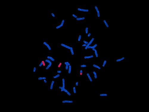
HOW DOES FISH TESTING WORK?

Step 1: A methanol-fixed blood smear is created from a patient’s blood sample.
Step 2: After adding the hybridization buffer with a pathogen-specific probe labeled with a fluorescent dye, the slide is incubated for a short time at 37℃. The pathogen rRNA will hybridize with the pathogen if present in the blood smear.
Step 3: After hybridization is complete, the excess probe is removed by washing with a special wash solution.
Step 4: The smear is dried, and counter-stain is added. The slide is then viewed with a fluorescent microscope; if pathogens are present, the blood smear will fluoresce.